Chapter 9: Genetics

Click here for Flashcards for this chapter
I. Section 1: Identifying the Genetic Material
A. Fredrick Griffith and Oswald Avery conduct Experiments that help to identify the existence of genetic material and its role in heredity.
1. Griffith wanted to create a vaccine to prevent pneumonia caused by the Streptococcus pneumonia bacterium.
a) He explored the reactions of mice to two types of the bacterium.
(1) S strain – smooth edged strain resulting from a capsule made of polysaccharides (causes the disease)
(2) R strain – rough edged strain lacking a capsule (does not cause disease)
b) Griffith assumed it was the capsule that was causing the disease
(1) First, he injected the mice with a form of the S strain that had been killed – the mice did not contract the disease
(2) Second, he injected the mice with a form of the S strain that had been heated and killed but left the capsule in tact – the mice still did not contract the disease – therefore it was concluded that it was not the capsule that caused the disease
(3) Third; he created a mixture of dead S strain and live R strain bacteria; the injected mice died; when he examined the blood of the dead mice he discovered that the harmless R bacteria had been transformed into the harmful S bacteria (the process of changing a phenotype by introducing foreign genetic material is now know as TRANSFORMATION)
2. Oswald Avery later added to these conclusions by determining that DNA was the material responsible for transformation; they learned that the instructions for making the bacteria were held in DNA code

B. Alfred Hershey and Martha Chase demonstrate that virus genes are also made of DNA

1. Scientists knew that viruses were made of DNA or RNA and a protein coat and that when a virus invaded a bacteria cell it was able to produce more bacteria
2. Hershey and Chase suggested that the virus would reprogram the bacteria itself to make the new viruses
3. They used a BACTERIOPHAGE ( DNA viruses that infect bacteria) test their ideas
a) Step One: They labeled the DNA of phages with radioactive Phosphorus-32; and labeled the protein coat of the phages with radioactive Sulfur-35
b) Step Two: They allowed the phages to infect E. coli (bacteria)
c) Step Three: They then removed the protein shells from the infected cells with a blender and a centrifuge. After separation, the radioactive tracer then was found in the protein shells, but not in the infected bacteria, confirming that the genetic material which infects the bacteria is DNA.
d) Step Four: It was determined that – upon infection the phages’ protein coat falls away; the phage then injects their DNA into the bacteria causing it to produce more viral DNA and proteins

II. Section 2: The Structure of DNA (deoxyribonucleic acid)
A. James Watson and Francis Crick determine the structure of DNA
1. Double Helix – two strands twisted around each other like a winding staircase or ladder
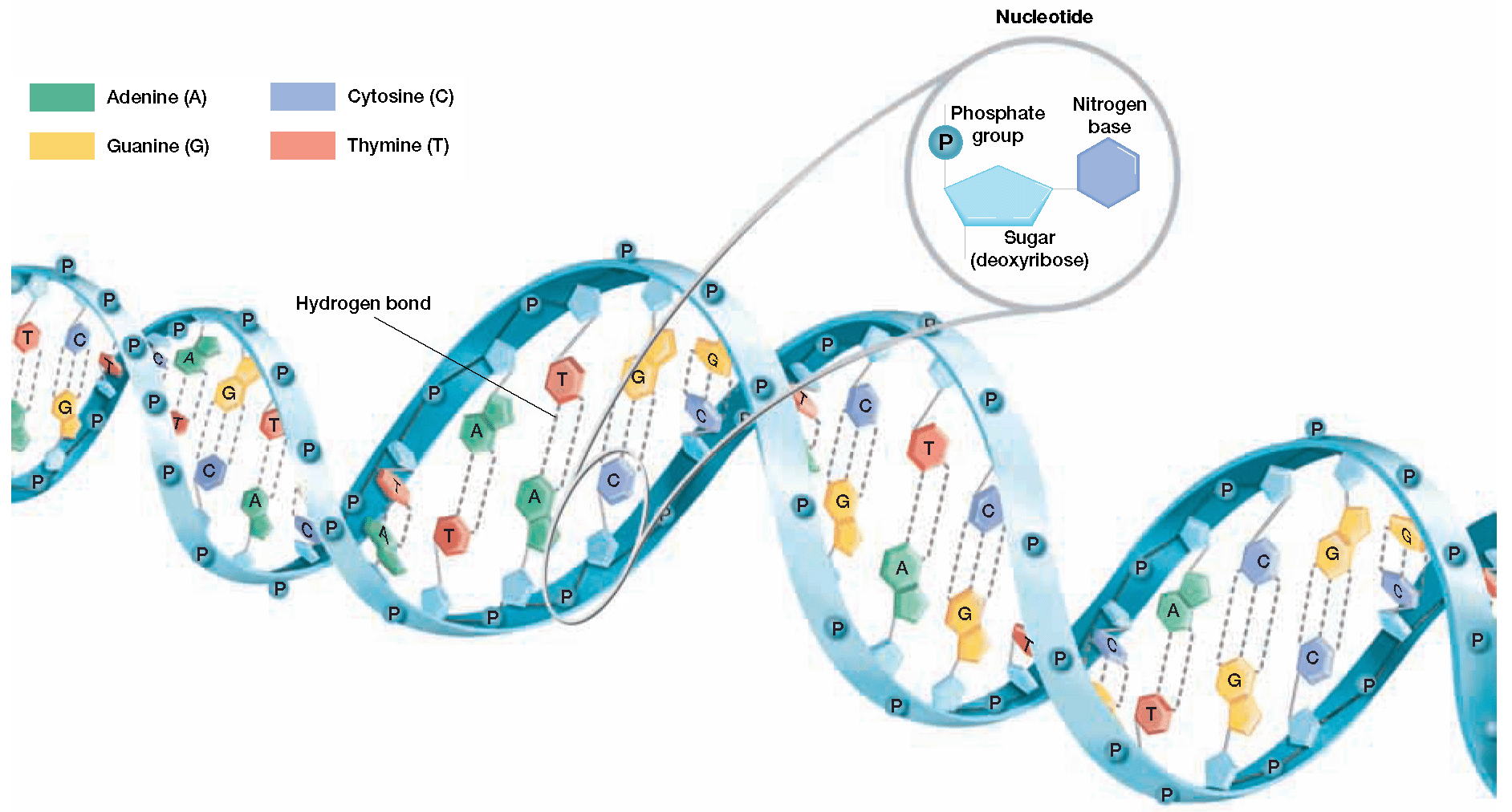
2. Nucleotides – subunits of DNA (each section of the ladder)
a) Three parts of the nucleotide; phosphate group, 5-carbon sugar, (these two make up the sides of the ladder) and a nitrogen-containing base (makes up the rungs of the ladder)

b) Four nucleotides exist in two groups
(1) The purines: adenine and guanine are bulky with double rings
(2) The pyrimidines: Thymine and Cytosine are smaller with single rings

III. Other Scientists Contributions
A. Erwin Chargaff
1. Discovered that the amount of cytosine always equaled the amount of guanine; the amount of thymine always equaled the amount of adenine

B. Maurice Wilkins and Rosalind Franklin
1. By analyzing the complex patterns on X-ray diffraction photo, scientists can determine the structure of the molecule.
2. In 1952, Maurice Wilkins and Rosalind Franklin developed high-quality X-ray diffraction photographs of strands of DNA.
3. These photographs suggested that the DNA molecule resembled a tightly coiled helix and was composed of two or three chains of nucleotides.
C. Watson and Crick develop a model of DNA
1. In 1953, Watson and Crick built a model of DNA with the configuration of a double helix, a “spiral staircase” of two strands of nucleotides twisting around a central axis.
2. The double-helical model of DNA takes into account Chargaff’s observations and the patterns on Franklin’s X-ray diffraction photographs
3. The model described pairing between bases (known as the base pairing rules); these rules are supported by Chargaff’s observations
(1) Cytosine always pairs with Guanine (forms 3 hydrogen bonds)
(2) Thymine always pairs with Adenine (forms 2 hydrogen bonds)
(3) The hydrogen bonds keep the two strands of DNA together and create strands that are complimentary to each other

IV. Section 3: The Replication of DNA
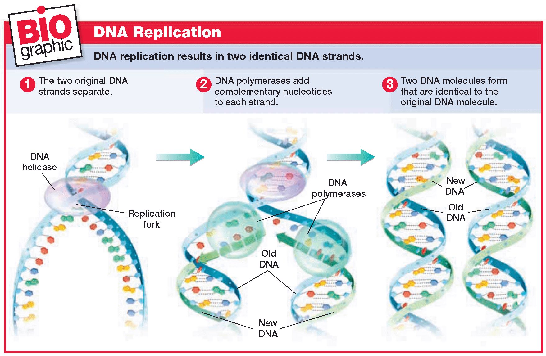
A. Role of enzymes in DNA replication (making a copy)
1. The complimentary structure of DNA is used to make exact copies of the DNA each time the cells divides
2. The process of making a copy of the DNA is called DNA replication
3. There are 3 steps in DNA replication
a) Step 1 DNA helicases open the double helix by breaking the hydrogen bonds that link the complementary nitrogen bases between the two strands. The areas where the double helix separates are called replication forks.

b) Step 2 At the replication fork, enzymes known as DNA polymerases move along each of the DNA strands. DNA polymerases add nucleotides to the exposed nitrogen bases, according to the base-pairing rules.
c) Step 3 Two DNA molecules form that are identical to the original DNA molecule
B. Checking for errors
1. In the course of DNA replication, errors sometimes occur and the wrong nucleotide is added to the new strand; this results in a mutation.
2. An important feature of DNA replication is that DNA polymerases have a “proofreading” role; the polymerase is able to erase the incorrect nucleotide and replace it with the correct one.
3. This proofreading reduces errors in DNA replication to about one error per 1 billion nucleotides.
C. Multiple replication “forks” increase the speed of replication
1. Replication does not begin at one end of the DNA molecule and end at the other.
2. The circular DNA molecules found in prokaryotes usually have two replication forks that begin at a single point.
